Real-time surgical tool segmentation from live operating-room footage.
A U-Net trained on 300 hand-labeled frames captured through a Modus V exoscope at Synaptive Medical and Robarts Research Institute, segmenting tools from tissue at ~95% DICE in under 0.12 s per frame.
1. Abstract
A convolutional segmentation network that traces surgical instruments out of background tissue, frame by frame, on real operating-room footage rather than phantoms or simulated images. Trained on 300 hand-labeled frames from a Modus V exoscope, then validated on a fully held-out surgical video across four lighting and tool-count scenarios.
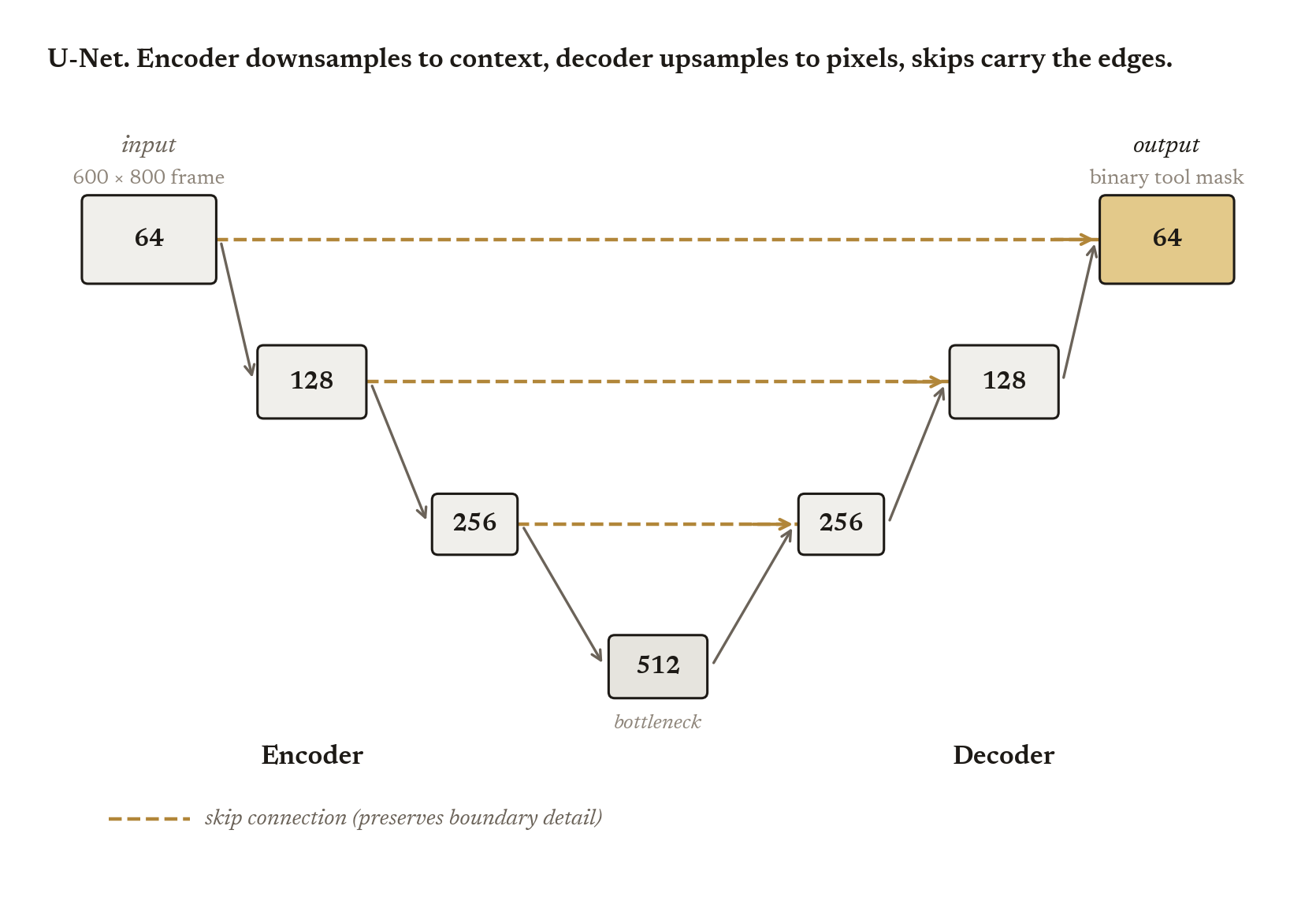
The model is a U-Net (Ronneberger et al., 2015), picked because its skip connections preserve the fine spatial detail that lands the tool boundary. Fluorescent-blue frames are converted to grayscale first: the network learns the tool more cleanly from intensity than from hue.
The novelty is not the architecture. It is the data, the partnership, and the deployment context: real exoscope footage on real tissue, not Cholec80 phantoms or synthetic renders; regular and fluorescence-guided imaging in one model; and a real-time inference loop on commodity hardware, demonstrated on video the network never saw.
- ~95% DICE on the canonical single-tool, regular-illumination case.
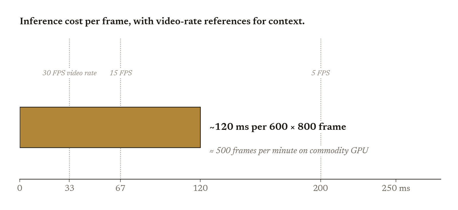
- Under 0.12 s per 600 × 800 frame, about 500 frames per minute on commodity GPU.
- 300 frames hand-segmented in ITK-SNAP, double-checked for ground-truth precision.
- Industry partnership with Synaptive Medical and Robarts Research Institute, captured on a Modus V exoscope.
- Bronze Medal, Canada-Wide Science Fair.
2. Introduction
Minimally invasive surgery asks the surgeon to operate through a keyhole. The camera replaces direct sight, depth perception collapses to a 2D feed, and the instrument tip routinely leaves and reenters the field. Existing literature attributes roughly 30% of complications in this setting to in-procedure misrecognition: tools confused with tissue, edges lost under fluorescence, occlusion read as background.
- The surgeon's view is already lossy. A real-time overlay that traces the tool boundary gives back information lost to camera distortion and shadow.
- Tool segmentation is the load-bearing primitive. Skill scoring, instrument tracking, autonomous assistance, and post-operative review all start from "where is the tool, pixel by pixel."
- The training set is the bottleneck, not the model. Public surgical-vision datasets skew toward laparoscopic or phantom footage.
2.1 Why a U-Net
U-Net was designed for exactly this regime: per-pixel classification on small biomedical datasets (300 frames, not 30,000), where the architecture has to compensate for what the data lacks.
- The skips land the tool boundary. They carry the high-frequency detail that pooling would discard. Without them the mask drifts several pixels off the actual edge; with them the boundary stays sharp, and the surgeon is looking at the edge, not the centroid.
- Tiny parameter count for a segmentation network. It trains end to end on a single GPU in roughly two hours, which kept iteration tight during the data and lighting experiments.
2.2 What is actually new
The architecture is from 2015. The contribution is the dataset, the lighting range, and the partnership that made both possible.
- 300 frames from a real Modus V exoscope on real tissue. Captured during experimental procedures on pig brain tissue, representative of operating-room lighting and focus. Not Cholec80. Not phantom data.
- Regular and fluorescence-guided imaging in one model. Grayscale conversion lets a single network handle both lighting modes without retraining (§3.2).
- Industry and clinical collaboration as part of the build. Synaptive Medical and Robarts Research Institute produced training data no public dataset has.
Everything below is engineering on top of those three. The architecture is the smallest piece.
3. System
3.1 Architecture
Standard U-Net. Three downsampling blocks (64, 128, 256 channels), a single 512-channel bottleneck, three upsampling blocks (256, 128, 64), and a final 1×1 convolution that produces the per-pixel tool probability. Skip connections concatenate encoder and decoder feature maps at matching resolution.
3.2 Data
300 frames sampled from the Modus V recording. Each frame was hand-segmented in ITK-SNAP into a binary tool / background mask, exported as NIfTI (for downstream tooling) and PNG (for training), and reviewed twice before entering the training set.
- Fluorescent-blue frames converted to grayscale in MATLAB. The blue channel carries the fluorescent signal but little tool-boundary information; intensity carries the boundary cleanly.
- Adam optimizer, binary cross-entropy loss.
- No augmentation beyond grayscale conversion on the fluorescent split. On a dataset this small, aggressive augmentation risked overfitting to synthetic artifacts rather than real intraoperative variation.
3.3 Test protocol
Evaluation runs on a fully held-out surgical video, captured separately and never seen in training. Frames are segmented individually and recompiled into a side-by-side overlay against the original footage. Four scenarios, 20 random images each:
- Regular illumination, one tool.
- Fluorescent blue, one tool.
- Regular illumination, two tools.
- Fluorescent blue, two tools.
The canonical case (regular, one tool) is the closest analogue to a standard intraoperative view and the headline number. The other three test how the model degrades away from it.
4. Evaluation
DICE on the binary mask is the quantitative metric; the side-by-side overlay is the qualitative check.
- ~95% DICE on the canonical single-tool, regular-illumination case.
- Under 0.12 s per 600 × 800 image: fast enough for surgical replay and near real-time overlay.
- Fluorescent blue reaches comparable accuracy to regular illumination on the single-tool case after grayscale conversion.
- Accuracy degrades on multi-tool frames, with fluorescent-blue two-tool the predicted worst case.
- Motion blur at tool edges is the primary failure mode, propagating into false positives and false negatives along the predicted mask edge.
The 95% number is the canonical case on n = 20 random frames from a single held-out video, single seed, single procedural source. It is not a leaderboard claim against published surgical-vision benchmarks. Read it as "this model works at video-realistic accuracy on data from this Modus V procedure," not as a SOTA result.
5. Conclusion and Implications
A real-time tool overlay trained on real surgical footage, rather than phantoms or synthetic renders, is a concrete step toward safer minimally invasive procedures. If an overlay that reliably traces tool boundaries recovers even a fraction of the 30% misrecognition share of intraoperative complications, the result is fewer nerve injuries, fewer collateral tissue insults, and fewer follow-on operations. Retrained on appropriate data, the same architecture extends to tumor delineation, organ boundaries for surgical planning, and quantitative tracking of disease progression on CT and MRI volumes.
5.1 Limitations and future work
- Single procedural source. All 300 frames come from one Modus V recording on one tissue type, so generalisation to other instruments, anatomies, and procedures is unproven. Next: a multi-source dataset under the same labeling protocol.
- Point estimate, hand-labeled ceiling. The 95% DICE is not variance-bounded, and ground truth is capped by manual ITK-SNAP labels despite two reviews per frame.
- Motion blur dominates the error. Each frame is segmented independently. Next: a recurrent or video-aware head to stabilise the mask across consecutive frames.
- Binary mask on multi-tool scenes. Next: instance segmentation with per-instrument labels for occluded multi-tool frames.
- Near real-time, not video rate. Live 30 FPS needs kernel-level optimisation, a leaner backbone, or quantised inference.
6. Acknowledgments
Surgical footage and procedural context provided by Synaptive Medical Inc., whose Modus V exoscope captured every frame in the training set. Tissue and clinical guidance provided by Robarts Research Institute at Western University. Recognized with a Bronze Medal at the Canada-Wide Science Fair.
The architecture is U-Net from Ronneberger et al., 2015.
@inproceedings{ronneberger2015unet,
  title     = {U-Net: Convolutional Networks for Biomedical Image Segmentation},
  author    = {Ronneberger, Olaf and Fischer, Philipp and Brox, Thomas},
  booktitle = {Medical Image Computing and Computer-Assisted Intervention (MICCAI)},
  year      = {2015},
  url       = {https://arxiv.org/abs/1505.04597}
}